Description
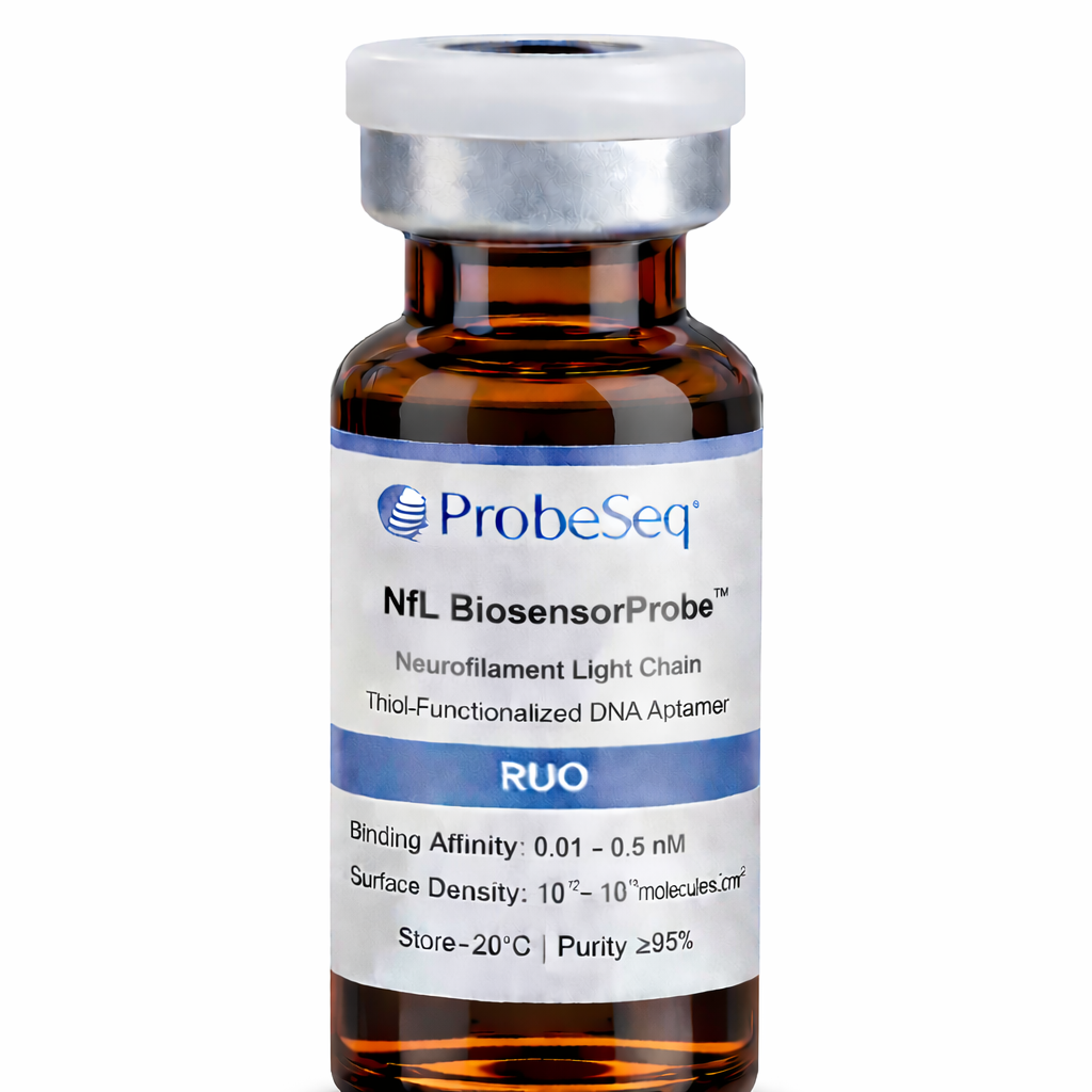
Surface-functional biosensor recognition ligand designed for selective detection of Neurofilament Light Chain (NfL), supplied as a thiol-modified DNA aptamer for direct immobilization on gold and thiol-reactive sensor surfaces. Engineered for high binding specificity, controlled probe orientation, and stable integration into electrochemical and microfluidic biosensor platforms. For Research Use Only (RUO).
Product Description
The ProbeSeq Neurofilament Light Chain BiosensorProbe™ is a thiol-functionalized single-stranded DNA aptamer engineered for selective recognition and capture of neurofilament light chain (NfL), a neuronal cytoskeletal protein widely used as a biomarker for neuroaxonal injury, neurodegeneration, and disease progression monitoring. The probe incorporates a terminal sulfhydryl (–SH) functional group enabling stable covalent immobilization on gold sensor interfaces while preserving aptamer folding and binding activity.
Neurofilament light chain is a structural protein (~68 kDa) released into cerebrospinal fluid and blood following neuronal damage. Baseline serum levels are typically <10 pg/mL and may increase to >100–1000 pg/mL in neurodegenerative disease and acute neurological injury. The BiosensorProbe™ supports ultrasensitive detection across clinically relevant concentration ranges.
The probe enables high-density surface immobilization (~10¹²–10¹³ molecules/cm²), controlled probe orientation, and stable signal generation across electrochemical, impedance, and optical sensing platforms.
Recognition Mechanism
Target recognition is mediated by a sequence-engineered single-stranded DNA aptamer (~45–85 nucleotides) forming a high-affinity binding structure specific to NfL epitopes through hydrogen bonding, electrostatic interactions, and structural complementarity.
Binding affinity (Kd): 0.01–0.5 nM
Detection range: pg/mL to ng/mL
Association kinetics: seconds to minutes
Reversible binding suitable for quantitative monitoring
Specificity and Selectivity
The NfL BiosensorProbe™ demonstrates high specificity toward neurofilament light chain with minimal cross-reactivity toward neurofilament heavy chain, tau protein, and GFAP.
Cross-reactivity: <3% against related neuronal proteins
Non-specific adsorption reduction: >90% vs non-functionalized DNA
Matrix compatibility: serum, plasma, and cerebrospinal fluid
Molecular Format and Functionalization
Synthetic single-stranded DNA aptamer containing terminal thiol functional group.
Aptamer length: ~45–85 nucleotides
Functionalization efficiency: ≥95% thiol incorporation
Purity: ≥95% (HPLC verified)
End modification: 5′ C6-thiol linker
Buffer formulation: nuclease-free buffered solution
Surface Immobilization Chemistry
Terminal thiol enables self-assembled monolayer formation on gold surfaces via Au–S bonding.
Surface binding strength: ~40–50 kcal/mol
Immobilization time: 30–120 min
Recommended surface density: 1–10 pmol/cm²
Operating pH after immobilization: 6.5–8.5
Compatible Sensor Surfaces
Gold electrodes and nanostructured gold substrates
Gold nanoparticle-modified interfaces
Thiol-reactive polymer coatings
Maleimide-functionalized surfaces
Electrochemical biosensors
Microfluidic diagnostic devices
Wearable neurological monitoring systems
Performance Characteristics
Binding affinity: 0.01–0.5 nM
Surface coverage: 10¹²–10¹³ molecules/cm²
Operating temperature: 4–45°C
Low non-specific adsorption
Stable signal response during repeated measurement cycles
Stability
Shelf stability: ≥12 months at −20°C
Working stability after immobilization: ≥14 days
Thermal tolerance: stable up to ~60°C short exposure
Storage
Store at −20°C in supplied buffer. Avoid repeated freeze–thaw cycles. Protect from oxidation and light.
Applications
Neurodegeneration monitoring
Multiple sclerosis biomarker detection
Alzheimer’s disease research
Traumatic brain injury detection
Electrochemical biosensor development
Microfluidic neurological diagnostics