Description
Product Overview
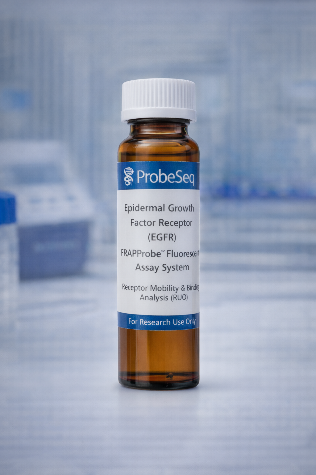
The ProbeSeq EGFR FRAPProbe™ System provides fluorescence-labeled probes for quantitative measurement of epidermal growth factor receptor (EGFR) mobility and binding kinetics in live cells and engineered tissue systems. The system enables characterization of receptor diffusion, clustering, trafficking, and ligand-induced signaling dynamics.
This product supports cancer biology research, therapeutic antibody screening, receptor–ligand interaction studies, and cell membrane transport analysis. The probe design ensures high photostability, controlled photobleaching behavior, and strong fluorescence recovery performance required for accurate FRAP measurements.
The system is compatible with confocal microscopy, live-cell imaging platforms, organ-on-chip devices, and microfluidic cell culture systems.
FRAP Technology Advantages
FRAP-based analysis enables quantitative measurement of molecular diffusion, receptor binding kinetics, and membrane transport dynamics in real time. Unlike endpoint biochemical assays, FRAP provides spatially resolved measurements of protein mobility within living systems. The approach allows direct evaluation of receptor trafficking, drug–target engagement, and membrane organization under physiological conditions. FRAP assays are particularly valuable for studying membrane receptor signaling, cancer biomarker behavior, and therapeutic response mechanisms.
Molecular Detection Mechanism
Fluorescently labeled EGFR probes bind specifically to EGFR expressed on the cell membrane. A defined region of interest is photobleached using a high-intensity laser pulse, followed by monitoring of fluorescence recovery as unbleached molecules diffuse into the region.
Quantitative recovery curves provide measurements of receptor diffusion coefficients, mobile fraction, and binding dynamics. The system supports analysis of receptor clustering, internalization, and ligand-induced signaling responses.
Target Design Strategy
Probe design targets extracellular domains of EGFR to enable selective receptor labeling while preserving native receptor function.
Design optimization prioritizes:
High binding specificity to EGFR
Minimal non-specific membrane interaction
Strong photostability under repeated imaging
Controlled photobleaching kinetics
Compatibility with live-cell imaging conditions
Stable fluorescence recovery behavior
Custom ligand-, antibody-, or nanobody-based probe formats are available.
Assay Workflow Integration
The FRAPProbe™ system supports flexible workflows including live-cell labeling, fluorescence imaging, targeted photobleaching, and recovery analysis.
The assay integrates with:
Confocal microscopy systems
High-content imaging platforms
Microfluidic cell culture devices
Organ-on-chip models
Drug screening platforms
Quantitative image analysis software
System Components
Fluorescent EGFR-binding probes
Recommended imaging and photobleaching parameters
Control reagents for calibration
Buffer and labeling guidelines
Data analysis recommendations
Custom probe formats available upon request.
Platform Compatibility
Confocal microscopy systems
Live-cell imaging platforms
High-content screening instruments
Microfluidic cell culture systems
Organ-on-chip devices
Drug screening workflows
Quantitative image analysis pipelines
Performance Characteristics
High fluorescence signal intensity, strong photostability, reproducible photobleaching response, rapid recovery measurement capability, low non-specific binding, and quantitative diffusion analysis performance. Performance depends on imaging configuration and cell model.
Technical Specifications
Detection target: Epidermal Growth Factor Receptor (EGFR)
Probe format: Fluorescent ligand / antibody / nanobody conjugate
Fluorophore options: GFP-compatible, Alexa Fluor, Cy dyes
Imaging compatibility: confocal FRAP systems
Working concentration: assay dependent
Storage: 4 °C or –20 °C depending on probe format
Quality Assurance
Binding specificity testing, fluorescence characterization, photostability validation, functional activity verification, and manufacturing quality review are performed for each batch.
Research Applications
Cancer biomarker mobility studies
Receptor signaling research
Drug–target engagement analysis
Therapeutic antibody screening
Membrane transport studies
Organ-on-chip receptor dynamics
Biosensor validation studies
Storage and Handling
Store under recommended temperature conditions protected from light. Avoid repeated freeze–thaw cycles. Use under sterile conditions for live-cell applications.